Transit through the Alimentary Canal (Digestive System)
Notice the difference
between the small diagram above and the larger diagram
on the right:
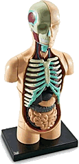
The diagram of
the digestive system above
indicates the locations
in the body of the tissues and organs of the digestive
system. It shows that they are packed together closely,
with some organs in front of, behind, above, and around
other organs. Remember that the reality (in 3-dimensions)
is more complicated that this 2-dimensional diagram,
which is further simplified by the use of hard lines
and shading different organs in different colours.
The diagram of
the digestive system on
the right is a schematic diagram,
meaning that the parts of the digestive system are
not really spaced-out as clearly as indicated in this
diagram. They are shown this way
to make it easier to see the route through the digestive
tract and so in which order each
part is reached.
Notice that ingested
materials do not pass through all of the organs
shown on the right.
Qu: Which parts does ingested material not
pass through, and what functions to those parts perform
?
The previous page identified the 4
basic stages of the digestive process.
This page describes in more detail the processes by
which foodstuffs (incl. ingested liquids) are broken
down and the useful parts taken into the cells of the
body. This includes further consideration of the organs
of the parts of the digestive system, whose locations
were indicated on the first page introducing
the human digestive system.
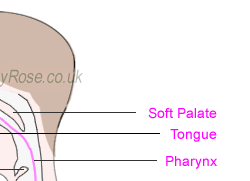
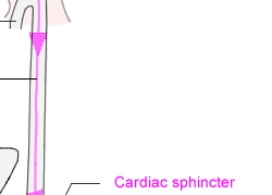
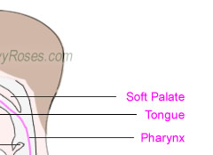
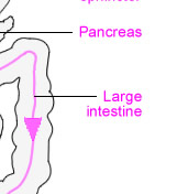
Above: Diagram of route through Alimentary Canal
Above: Diagram of route through Alimentary Canal
Notes about each stage of the alimentary canal:
The following are key points about the stages mentioned - with just sufficient extra detail to describe and explain the passage of ingested material through each part of the alimentary tract. These short points are in the form that might be required in a test or exam. For more about individual tissues and organs, use the links to further anatomical details (included in the text below).
1. |
Buccal Cavity |
Buccal Cavity is a medical term used
to refer to the mouth.
Several digestive processes occur in the buccal
cavity, including:
- Mechanical parts of the digestive process
include chewing and grinding using
the teeth.
- Saliva is produced and secreted by the salivary glands.
The secretion of saliva by the salivary glands is called salivation.
The production and secretion of saliva is increased
in response to the chewing action of the jaws,
and also in response to the thought, taste,
smell, and hence the experience of ingesting
foods. Saliva has several functions, including
lubrication of the buccal cavity.
- Saliva includes the enzyme salivary
amylase, which begins the process
of breaking-down the carbohydrates within the
food. Salivary amylase has the effect of breaking
down certain large molecules: polysaccharides
 di-saccharides. di-saccharides.
- Finally, after reduction to an appropriate
size and consistency, food is formed into a
"bolus" (i.e. a "ball" of
foodstuff) to be passed down the digestive tract.
|
2. |
Epiglottis |
The epiglottis is a thin leaf-shaped flap of cartilage covered with a layer of mucous membrane. It is
located immediately behind the root of the tongue
and aids the digestive process by closing the
trachea (which is sometimes colloquially referred
to as the "windpipe") to prevent
ingested materials from entering the lungs / respiratory
system. |
3. |
Trachea |
The trachea (or, colloquially, the "windpipe") is the part of the air passage between the larynx (which is sometimes colloquially referred to as
the "voice box") and the main
bronchi inside the lungs. Although not part of the digestive system, the trachea is labeled
above to indicate the importance of the epiglottis. |
|
4. |
Oesophagus |
The oesophagus (also known colloquially as the "windpipe") is the tube through which a bolus is carried from the mouth to the stomach. The bolus progresses down the oesophagus by means of peristaltic action. |
5. |
Cardiac Sphincter |
The cardiac sphincter is the site at which material enters the stomach.
A bolus passes from the oesophagus into the stomach through the cardiac sphincter. |
6. |
Stomach |
The stomach is a muscular sac that churns and mixes food. It also absorbs alcohol.
Mucus and proteases are
also present in the stomach.
Key Notes about the stomach:
- churns / mixes food
- mixes food with gastric acid
Enzymes in the stomach:
- pH approx. 1-3 due to stomach acid.
- Kill microbes.
- Neutralise salivary amylase.
- Provide a medium for proteases such as rennin
(coagulates milk proteins) and pepsin.
A protese is any enzyme that catalyses the splitting
of a protein.
Proteases catalyse the process: Polypeptides  Di-peptides Di-peptides  Amino acids. Amino acids.
|
7. |
Pylonic Sphincter |
The pylonic sphincter is the route by which
material exits the stomach.
The contents of the stomach is squeezed out as chyme into the small
intestine. |
8. |
Liver |
The liver is an accessory organ (i.e. it assists
the digestive process, e.g. by supplying substances
useful to the digestive process - but ingested
material does not pass through the liver).
The liver is the largest organ in the body,
the skin being the largest organ of the
body.
The liver has over 500 functions, including:
- Production and secretion of bile and bile
salts.
(Bile = blood pigments from erythrocytes + bile
salts + cholesterol). Bile is alkaline and its
function is to break-down ("emulsify"
= "make into smaller globules") fats.
- Phagocytosis of bacteria and dead or foreign
materials.
- Converts glucose to glycogen and vice-versa.
- Production of cholesterol
- Storage of glycogen
- De-amination of excess amino acids
- Detoxification, e.g. conversion of ammonia
to urea, and processing of alcohol and/or drugs.
- Storage of certain vitamins & minerals,
e.g. iron (Fe) that can be used to produce red
blood cells.
For more about these see the page about functions
of the liver. |
|
9. |
Pancreas |
The pancreas is an accessory organ (i.e. it assists the digestive process, e.g. by supplying substances useful to the digestive process - but ingested material does not pass through the pancreas). The pancreas is a "dual organ", i.e. it is both exocrine
and endocrine.
Endocrine Functions: Produces the hormones insulin and glucogon, which control sugar levels.
Exocrine Functions: Produces enzymes:
- Pancreatic Amylase - breaks down carbohydrates by:
Polysaccharides  Di-saccharides Di-saccharides
- Lipase - breaks down fats by:
Fat  Fatty Acids + Glycerol Fatty Acids + Glycerol
- Proteases e.g. typsin - break-down proteins by:
Polypeptides  Di-peptides Di-peptides  Amino acids Amino acids
|
10. |
Small Intestine |
There are three parts of the small intestine.
They are (in the order in which they are reached):
- The Duodenum
- The Jejunum
- The Ileum
In addition to the enzymes already contributed at previous stages in the alimentary tract, further enzymes are released from the walls of parts (1.) and (2.) of the small intestines.
Examples include: sucrase, maltase, fructase, lactase.
The '-ase' suffix indicates that the substance is an enzyme.
These facilitate reactions of the form: Di-saccharides  mono-saccharides, mono-saccharides,
so, e.g.
The enzyme sucrase facilitates
break-down to the mono-saccharide sucrose.
The enzyme maltase facilitates
break-down to the mono-saccharide maltose.
The enzyme fructase facilitates break-down to the mono-saccharide lactose.
The enzyme lactase facilitates
break-down to the mono-saccharide lactose.
Absorption takes place at (3.), then ...
- Glucose and amino acids go to the hepatic portal vein, and
- Fatty acids and glycerol go into lacteal and are transported by the lymphatic system.
|
11. |
Ileocaecal Valve |
The iloecaecal valve is the exit through which chyme passes from the small intestine to the large intestine. |
12. |
Appendix |
Note
that the appendix is not strictly part of the
alimentary tract. It is mentioned here to complete
brief notes about all of the tissues and organs
labeled in the diagram above.
The appendix is a "vestigial organ", which means
that it is thought to be present in the body as
a result of evolution - even though the human
body has evolved in such as way as to render it
(the appendix)
non-essential.
Ingested matter does not pass through the
appendix.
The appendix is composed of lymphatic
tissue. |
13. |
Large Intestine |
The large intestine is the final organ in the alimentary tract.
It consists of sections that have specific names, including:
The large intestine absorbs water from material passing through it all the way along its length.
The final stages of the large intestine (the rectum and anal canal) also form and release faeces, as stated below. |
|
14. |
Rectum |
The rectum is a latter part of the large intestine. Its purpose is the formation of faeces, i.e. faeces are formed in the rectum then expelled via the process of defecation. |
15. |
Anus |
The anus is the opening at the lower-end of the alimentary
tract, through which faeces are discharged.
The anus opens out from the anal canal (which is the end, or "terminal", portion
of the large intestine) and is kept closed by
two sphincter muscles at all times except during defecation. |
 |
 |
 |
|
The above introduces the components of the alimentary tract and presents key points about the tissues and organs through which ingested matter (i.e. foodstuffs) pass during the digestive process.
The key-points listed in note form above may not include sufficient information even for introductory-level courses that include human digestion. For more about the anatomy and physiology of individual parts of the alimentary tract, see the pages about the teeth, oesophagus, stomach, liver, pancreas, small intestine and large intestine.
The next page in this series is about the structures of the mouth.


