
Functions of Microtubules in Cells
Main Functions of Microtubules:
- Microtubules are the main "building blocks" that form the cytoskeleton - which is the cell's framework within which all components of the cell are held in position or allowed to move within certain constraints.
- Movement of materials and structures within cells.
e.g. they help form the miotic spindle apparatus via which chromosomes move to two poles during cell division by mitosis. This occurs during the "prophase" part of mitosis - see diagram below.
See below for further information ...
Re-cap: What are microtubules ?
Microtubules are present in eukaryotic cells, including
- plant cells
and - animal cells.
Microtubules are long thin structures that consist of the protein tubulin and typically have a diameter of about 25 nm. Characteristics of microtubules that are important for their functions include:
- Long rigid shape - which enables microtubules to support other structures within the cell
- Ability to generate movement - both within cells (see their role in moving centrioles during mitosis - below) and of the whole cells themselves (in the cases of microtubules that form structures such as cilia and flagella).

More about the structure of microtubules
Microtubules consist of longitudinally arranged assemblies of filaments called protofilaments. These protofilaments group together to form long cylinders or tubes called microtubules such that a cross-sectional view of a microtubule would look like a circle formed by the cross-sections (diameters) of approximately thirteen (13) protofilaments.
The protofilaments themselves have two forms - they are both molecules of the protein tubulin but they differ in the sequence in which the amino acids forming the protein are arranged. The two types of protofilaments are:
- alpha tubulin (sometimes written α-tubulin)
and - beta tubulin (sometimes written β-tubulin)
1. Microtubules are an important part of the cytoskeleton of cells
Electron microscopy has revealed that cells contain a network of interconnected fibres and filaments that can be observed as thread-like structures when viewed using an electron microscope or fluorescence microscope (tagged with antibodies and fluorescent dyes). This network is called the cytoskeleton. It extends throughout the cell and connects with the cell membrane and organelles.
The main functions of the cytoskeleton of a cell are: |
The main components of the cytoskeleton of cell are: |
|
|---|---|---|
|
|
2. Role of microtubules during the prophase stage of mitosis
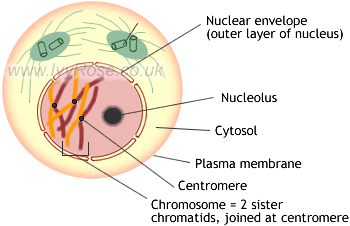
The two diagrams below illustrate the prophase stage of mitosis in which the following occurs:
- Early in the prophase stage the chromatin fibres shorten into chromosomes that are visible under a light microscope. (Each prophase chromosome consists of a pair of identical double-stranded chromatids.)
- Later in prophase, the nucleolus disappears, the nuclear envelope breaks down, and the two centrosomes begin to form the miotic spindle (which is an assembly of microtubules).
- As the microtubules extend in length between the centrosomes, the centrosomes are pushed to opposite "poles" (extremes) of the cell.
- Eventually, the spindle extends between two opposite poles of the cell, as shown below.
 |
 |
|
|---|---|---|
Above: Early Prophase |
Above: Late Prophase |
The mitotic spindle is formed during prophase and remains within the dividing cell, an important part of its structure, through metaphase and anaphase until it finally breaks-up when the two new cells separate during telophase. See diagrams of mitosis.
Remember : A microtubule is found inside some cells but is not a (type of ) cell.
For more about cells: the structure of a plant cell, the structure of an animal cell & comparison of plant, animal and bacterial cells. See also very simple information about chloroplasts and photosynthesis (via 3 short YouTube videos).


