Anatomy of The Eye
The anatomy and physiology of the human eye is an important part of many courses e.g. in biology, human biology, physics, and practical courses in medicine, nursing, and therapies.
This simple introduction the subjects of 'the eye' and 'visual optics' includes a simple diagram of the eye together with definitions of the parts of the eye labelled in the illustration.
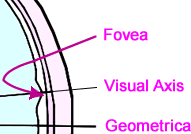
Above: Schematic diagram of the Structure of the Human Eye.
Term |
Definition / Description |
|
Aqueous Humour |
The aqueous humour is a jelly-like substance located in the anterior chamber of the eye. |
... more about the Aqueous Humour |
Choroid |
The choroid layer is located behind the retina and absorbs unused radiation. |
... more about the Choroid |
Ciliary Muscle |
The ciliary muscle is a ring-shaped muscle attached to the iris.
It is important because contraction and relaxation of the ciliary muscle controls the shape of the lens. |
... more about the Ciliary Muscle |
Cornea |
The cornea is a strong clear
bulge located at the front of the eye (where
it replaces the sclera - that forms the
outside surface of the rest of the eye).
The front surface of the adult cornea has
a radius of approximately 8mm.
The cornea contributes to the image-forming
process by refracting light entering the
eye. |
... more about the Cornea |
Fovea |
The fovea is a small depression
(approx. 1.5 mm in diameter) in the retina.
This is the part of the retina in which
high-resolution vision of fine detail is
possible. |
... more about the Fovea |
Hyaloid |
The hyaloid diaphragm divides
the aqueous humour from the vitreous humour. |
... more about the Hyaloid Membrane |
Iris |
The iris is a diaphragm
of variable size whose function is to adjust
the size of the pupil to regulate the amount
of light admitted into the eye.
The iris is the coloured part of the eye
(illustrated in blue above but in nature
may be any of many shades of blue, green,
brown, hazel, or grey). |
... more about the Iris |
Lens |
The lens of the eye is a
flexible unit that consists of layers of
tissue enclosed in a tough capsule. It is
suspended from the ciliary muscles by the
zonule fibers. |
... more about the Lens |
Optic Nerve |
The optic nerve is the second
cranial nerve and is responsible for vision.
Each nerve contains approx. one million
fibres transmitting information from the
rod and cone cells of the retina. |
... more about the Optic Nerve |
Papilla |
The papilla is also known
as the "blind spot" and is located
at the position from which the optic nerve
leaves the retina. |
... more about the Optic Papilla |
Pupil |
The pupil is the aperture
through which light - and hence the images
we "see" and "perceive"
- enters the eye. This is formed by the
iris. As the size of the iris increases
(or decreases) the size of the pupil decreases
(or increases) correspondingly. |
... more about the Pupil |
Retina |
The retina may be described
as the "screen" on which an image
is formed by light that has passed into
the eye via the cornea, aqueous humour,
pupil, lens, then the hyaloid and finally
the vitreous humour before reaching the
retina.
The retina contains photosensitive elements
(called rods
and cones)
that convert the light they detect into
nerve impulses that are then sent onto the
brain along the optic nerve. |
... more about the Retina |
Sclera |
The sclera is a tough white
sheath around the outside of the eye-ball.
This is the part of the eye that is referred
to by the colloquial terms "white of
the eye". |
... more about the Sclera |
Visual Axis |
A simple definition of the
"visual axis" is "a straight
line that passes through both the centre
of the pupil and the centre of the fovea".
However, there is also a stricter definition
(in terms of nodal points) which is important
for specialists in optics and related subjects. |
... more about the Visual Axis |
Vitreous Humour |
The vitreous humour (also
known as the "vitreous body")
is a jelly-like substance. |
... more about the Vitreous Humour |
Zonules |
The zonules (or "zonule
fibers") attach the lens to the ciliary
muscles. |
... more about the Zonules |
|
To learn more about the eye and how it works it is useful to understand some simple but important aspects of light.
Next: Understanding Light (to explain how the eye works).

